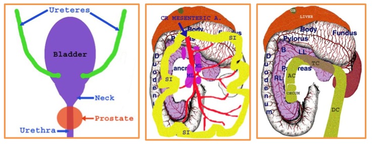
How To Ultrasound the Abdomen

Dr. Rendano's Protocol for Examining the Abdomen
  • Place the pet in dorsal recumbency if appropriate; consider sedation if the pet will not stay and if sedation is appropriate.


  • Clip hair from the ventral abdomen if needed and if permitted – sternum to pubis and laterally as far as needed to be able to visualize the kidneys, right adrenal gland and spleen.


  • Wet the area to be imaged with alcohol and apply coupling gel.


  • Place appropriate frequency probe/transducer on the abdomen to visualize the spleen and left kidney-refine power, gain, TGC settings etc. as needed.


  • Each organ system should be evaluated in sagittal and transverse planes.
    •  Static images can be saved and organs/lesions measured as appropriate.


  • Start recording the study and proceed as follows-clockwise assessment the abdominal viscera:


  • Place probe by last sternebra and then move it to evaluate:
    • left side of liver
    • right side of liver
    • gall bladder
    • portal vein andporta hepatis
    • spleen
    • left kidney
    • left adrenal gland
    • urinarybladder
    • reproductiveorgan(s)
    • sublumbar lymph nodes
    • right kidney
    • right adrenal gland
    • stomach
    • bowel
    • mesenteric lymph nodes
    • pancreas
abdomen 1

abdomen 3
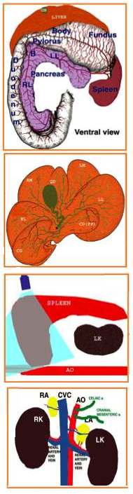
  • Place animal in right lateral recumbency and evaluate:
    • pancreas (if not previously seen)
    • left adrenal gland (if not previously seen


  • Place animal in left lateral recumbency and evaluate:
    • right adrenal gland (if not previously seen)


  • Make concluding statements after you evaluate each organ system. If there is a mass lesion define, freeze, label and measure it.